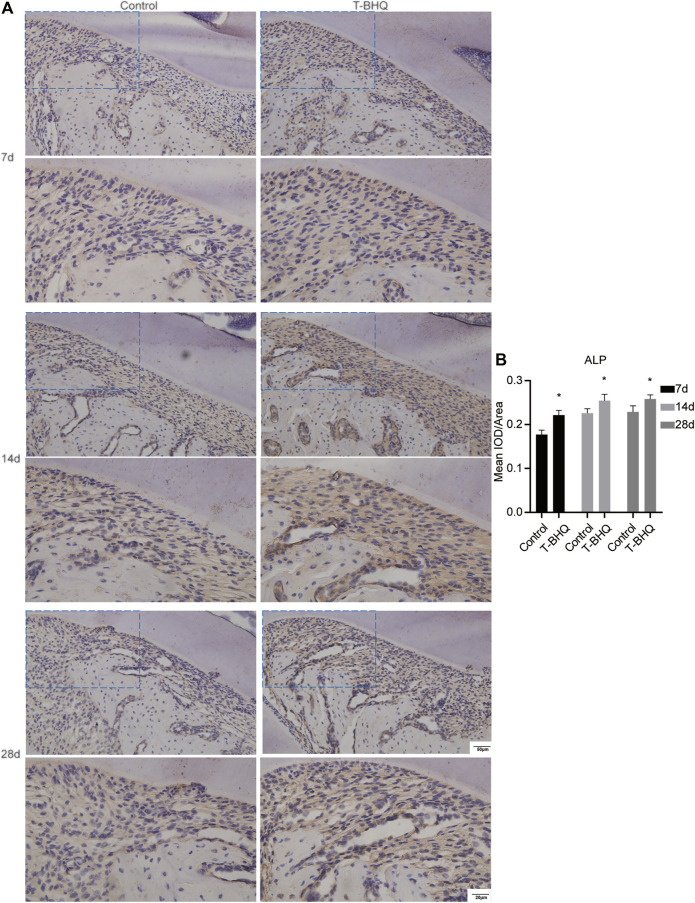
FIGURE 7.
The increased expression of ALP in the PDL at the tension side in orthodontic rats with t-BHQ treatment. (A) The expression of ALP was enhanced with t-BHQ treatment in the PDL at the tension side in orthodontic rats through IHC staining. The scale bars, 20 and 50 µm. (B) The mean IOD/Area of ALP was quantified through Image-Pro Plus 6.0 software. N = 3 specimens in each group.

