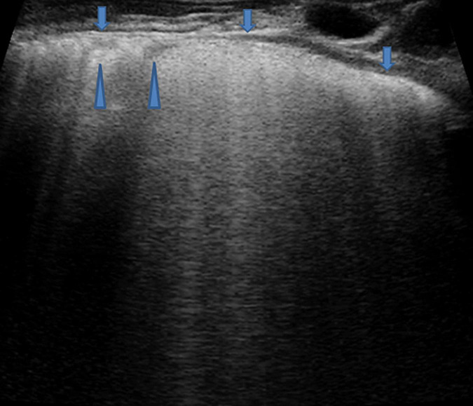
FIGURE 9.
Grayscale lung ultrasound examination (transverse scan between intercostal fields; linear probe with 12 MHz frequency) of a 4-year-old boy with viral pneumonia – due to Coronavirus (non-COVID-19), Bocavirus, and Metapneumovirus coinfection- requiring respiratory assistance with High- flow nasal oxygen at the pediatric department. It shows sonographic interstitial syndrome (SIS) which is characterized by blurred, uneven, coalescent B-lines and white lung; irregular pleural line (arrows); reduced pleural sliding; multifocal inhomogeneous involvement; subpleural microconsolidations (generating pseudo-B-lines) (arrowheads).

