Figure 2.
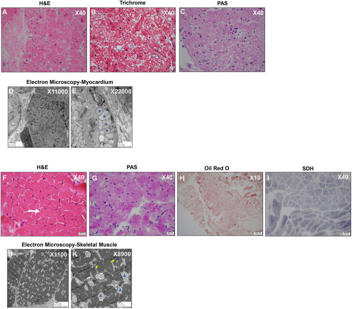
Myocardium and Skeletal Muscle Biopsies Reveal Hyperplastic and Polymorphic Mitochondria and Lipid Accumulation. (A) Myocardium: H&E X40 shows diffuse generalized granular vacuolization likely representing mitochondria. (B) Myocardium: Mason Trichrome X40 reveals minimal fibrosis. (C) Myocardium: Periodic Acid Schiff (PAS) X40 Negative staining indicates absence of abnormal glycogen content. (D,E) Myocardium: Electron microscopy– myocardium X11000 (D) and X22000 (E): Mitochondrial hyperplasia, pleoconia, and megaconial forms and loss of internal cristae; Stars indicate mitochondria of variable and abnormal size. (F) Skeletal Muscle: H&E X40 Moderate fiber size variation with occasional atrophic fibers (white arrow). No evidence of degeneration/regeneration/inflammation. (G) Skeletal Muscle: PAS X40 Mild glycogen accumulation. (H) Skeletal Muscle: Oil Red O X10 Increased cytoplasmic lipid. (I) Skeletal Muscle: SDH X40 complex II mitochondrial respiratory chain enzyme-normal appearance. (J,K) Electron microscopy-skeletal muscle direct magnification of X11000 (J) and X8900 (K): Mitochondria are normal in size and shape (yellow arrows); there is slight increase in cytoplasmic Glycogen (orange star) lipid (blue arrowhead).

