
Figure 2.
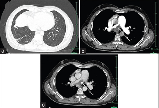
(a) High-resolution computed tomography lungs shows juxtapleural fibrosis, suggestive of postinfective changes in the middle lobe of the right lung. (b) (Thin arterial postcontrast pulmonary angiography) - shows a right main pulmonary artery thrombus (arrow) causing a narrowing of the vessels downstream. (c) (CTPA, delayed image)-shows similar findings