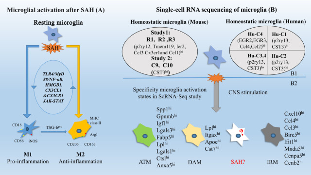
Figure 1.
Microglial activation states.
The left part shows the activation of microglia after subarachnoid hemorrhage (SAH) (A). M1 (Marker CD16, CD86; iNOS) have been observed to have the proinflammatory effect and M2 (Marker CD206, CD163, Arg1 and MHC class II) have an anti-inflammatory effect by interacting with signal pathway TLR4/MyD88/NF-κB, HMGB1, CX3CL1 &CX3CR1 and JAK-STAT. The right part (B) shows the latest results of single-cell RNA sequencing study on transcriptional states of homeostatic microglia (B1) and activated microglia (B2). Two to three clusters of resting microglia in mice were found and with an expression of homeostatic genes. Healthy human microglia were found to have four clusters: Human Cluster 1 (Hu-C1) to human cluster 4 (Hu-C4). Each microglia sub-state has its own up-regulated gene and some clusters have several similar characteristics. The activated microglia have also been analyzed in a single-cell study. DAM was a subcluster of activated microglia found in Alzheimer's disease mouse model. Specialized Axon Tract-Associated Microglia (ATM) were observed in the developmental mouse brain. IRM is another subtype of activated microglia observed in lysolecithin induced animal model. DAM, ATM and IRM all shared a common transcriptional signature of 12 core genes including Spp1, Lpl, and Apoe. There were also differential gene expressions among them. However, microglia transcription heterogeneity leaves unknown in the SAH state.

