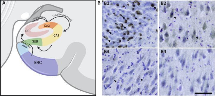
Figure 1.

(A) Information from layer II neurons in the entorhinal cortex (ERC) travels to the granule cells of the dentate gyrus (DG) via the perforant pathway (Geula, 1998).
Mossy fiber axons from the DG granule cells then carry information to CA3, and Schaffer axon collaterals project to the CA1 (Jamshidi et al., 2020). Finally, information from CA1 travels to the subiculum. (B) Hippocampal distribution of TDP-43 mature inclusions and pre-inclusions. (B1) Granule cells of the dentate gyrus show a high density of both mature inclusions (arrows) and pre-inclusions (arrowheads). (B2) CA3 pyramidal cells demonstrate a high abundance of pre-inclusions (arrows) but scarce mature inclusions. (B3) The CA2 subfield shows relatively fewer pre-inclusions compared to the CA3 subfield and a similar absence of mature inclusions. (B4) In the CA1 subfield, pre-inclusions and mature inclusions are virtually absent. Photomicrographs were acquired using 40 μm hippocampal sections stained with a human phosphorylated TDP-43 antibody and counterstained for Nissl using Cresyl violet. Scale bar: 100 μm.
