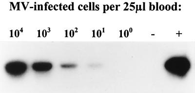
FIG. 1.
RT-PCR detection of in vitro MV-infected cells diluted in human blood and spotted on filter paper. A human Epstein-Barr virus-transformed B-LCL was infected with a wild-type MV isolate from Khartoum, washed, counted, diluted in human blood with EDTA, and spotted on filter paper in 25-μl samples. After storage of the filter paper samples at room temperature for 6 weeks, RNA was isolated and RT-PCR was carried out with primers MV-N1 and MV-N2. The resulting amplicons were of the correct size as estimated on the gel using a 100-bp ladder as reference (not shown). The PCR products were blotted and hybridized with 32P-labeled oligonucleotide probe MV-prN2. The autoradiagram is shown, with numbers of MV-infected cells per 25 μl indicated above the respective lanes. The positive (MV Edmonston) and negative (untreated human blood with EDTA) controls are indicated by + and −, respectively.

