Figure 1.

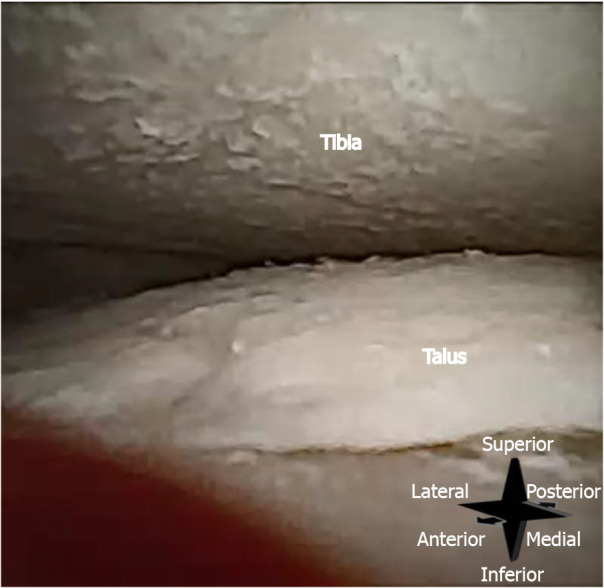
Intra-articular image of a right tibiotalar joint, taken with the 0° arthroscope inserted through the anteromedial portal. Substantial chondral wear can be seen on talus and tibia, with uncovered bone clearly visible.

Intra-articular image of a right tibiotalar joint, taken with the 0° arthroscope inserted through the anteromedial portal. Substantial chondral wear can be seen on talus and tibia, with uncovered bone clearly visible.