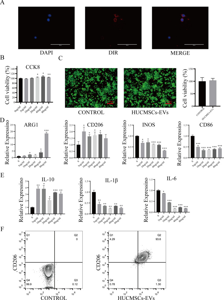
Fig.3.
HUCMSCs-EVs effectively promote the polarization of M2 macrophages in vitro. A hUCMSCs-EVs were labeled with DIR, and uptake of DIR-labeled hUCMSCs-EVs by macrophages was observed by confocal microscope; Scale bar: 100 μm. B The proliferative effect of hUCMSCs-EVs on macrophages was measured by CCK-8 analysis. C The impact of hUCMSCs-EVs on the viability of macrophages was detected by the cell live/death experiment; green represents live cells while red represents dead cells; Scale bar: 1 mm. D Relative mRNA expression of the critical genes ARG1, CD206, INOS, and CD86 was determined in polarized macrophages by quantitative RT-PCR analysis; the data of triplicate experiments are presented as mean ± S.D. *p < 0.05, **p < 0.01, ***p < 0.001. E Relative mRNA expression of the related cytokines IL-10, IL-1, and IL-6 were determined in polarized macrophages by quantitative RT-PCR analysis; the experiment was conducted triplicate; *p < 0.05, **p < 0.01, ***p < 0.001. F The typical surface markers in M2-polarized macrophages induced by hUCMSCs-EVs were detected by Flow cytometry analysis

