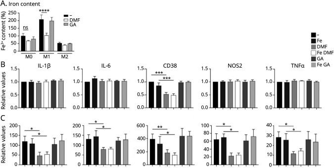
Figure 3. Changes in Iron Content and Activation State of Human Microglial Cell Cultures.
(A) Iron content in human iPSC-derived, M0-, M1-, and M2-polarized microglial cells was quantified with intracellular radioactive 55Fe after treatment with/without DMF and GA. Quantification of mRNA expression of IL-1β, IL-6, TNF, CD38, and NOS2 in (B) unpolarized and (C) M1-polarized microglia, incubated with and without FeCl3 and treated with DMF or GA. Data are normalized to the untreated, unpolarized microglia and represent mean ± SD. *p < 0.05, **p < 0.01, ***p < 0.005, and ****p < 0.0001; ns = nonsignificant. DMF = dimethyl fumarate; GA = glatiramer acetate; IL = interleukin; iPSC = induced pluripotent stem cell; mRNA = messenger RNA; TNF = tumor necrosis factor.

