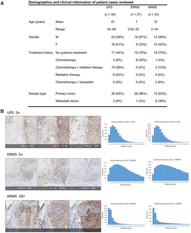
Figure 2.
A, Patient demographics and clinical history of all cases reviewed. B, Three representative cases illustrating tumor vasculature and immune cell infiltration. IHC slides stained with CD3 (T cells), CD163 (TAMs), and CD31 (endothelial cells) provide a geographic overview of UPS, ERMS, and ARMS specimens (left). In all three subtypes, the majority of T cells and TAMs cluster near endothelial cells. Corresponding histograms represent proximity analysis with HALO pathology software measuring distance (mm) between CD3+ cells and CD31+ cells, as well as between CD163+ cells and CD31+ cells (right). Results demonstrate the majority of T cells and TAMs to be found within 40 μm to tumor endothelial cells in UPS, ERMS, and ARMS.

