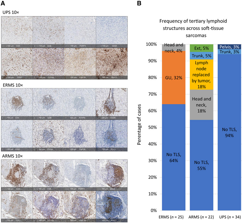
Figure 4.
Organization of immune cells and frequency of TLS. A, IHC stains shows diffuse distribution of T cells (CD3+ and CD8+) in UPS with no B cells (CD20+) present. In ERMS and ARMS, T cells (CD3+ and CD8+) cluster together with B cells (CD20+) forming TLS. TAMS (CD163+) are diffusely distributed in all sarcomas. CSF1R appears stronger in UPS and ARMS. PD-L1 is present through all sarcomas but stronger in UPS. B, The frequency of TLS in each sarcoma subtype varies and originates from different anatomical sites.

