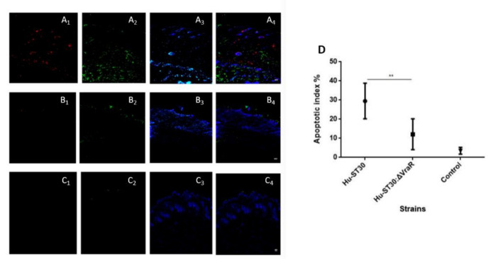
Figure 7.
Comparison of MRSA ST30 and ST30ΔvraR using skin and soft-tissue infection (SSTI) model. Mice were inoculated subcutaneously with either PBS or bacteria as described in Methods. (A) A double-labeling assay for detection of the staphylococcal invasion of the skin model with anti-Staphylococcus aureus antibody and detection of the apoptotic cells, using TUNEL assay after 48 h of infection with the MRSA. (A) ST30 and (B) ST30ΔvraR, and (C) PBS (1) anti-Staphylococcus aureus antibody with goat anti-rabbit IgG H and L conjugated with Alexa Fluor 568 secondary antibody, (2) the Click-iT® TUNEL Alexa Fluor® 488 cells (3) Hoecsht stain, and (4) the overlay of the emission signals. White hashed line demarcates the dermal epidermal boundary between the strata basale and the collagen gel populated with fibroblasts. Scale bar shows 50 µm. (D) Apoptotic index. Ten fields were calculated for each condition and original magnifications of images are 100×. Data are shown as the means ± SD, significance were determinant by one-way ANOVA and coded as ** p < 0.01.

