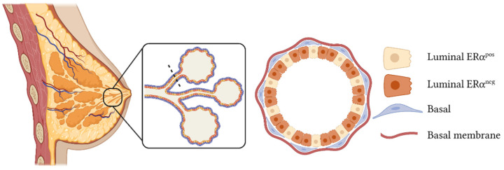
Figure 1.
Model of normal mammary gland structure. This tissue is composed of ducts, which are formed by three epithelial populations: basal cells, in contact with the basal membrane; estrogen receptor-positive (ERαpos) luminal cells; and estrogen receptor-negative (ERαneg) luminal cells. The dotted black line indicates the cross-section of the mammary duct represented in the magnified scheme on the right. This figure was created with Biorender.com (accessed on 12 December 2021).

