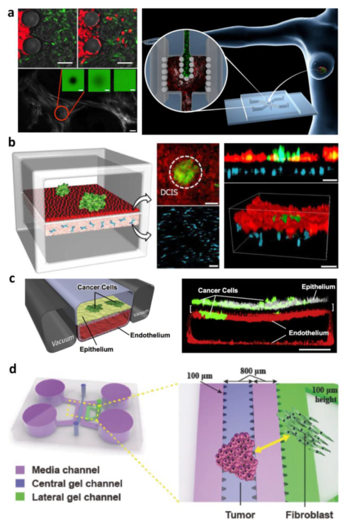
Figure 3.

Cancer on chip for modeling tumor–stroma interaction: (a) On-chip activation of stromal tissue by crosstalk with cancerous tissue. The compartmentalized device is designed for accommodating stromal tissue and epithelial tumor tissue allowing microtissues physical contact, across the separation formed by the pillars, in order to replicate the tissue–tissue interface. SHG and FRAP techniques were used to investigate transport properties and remodeling of neo synthesized ECM and time-lapse images at fluorescence microscopy were used to detect the migration of MCF7 cells from the tumoral chamber to the stromal chamber. Reproduced with permission [92]. Copyright 2016, John Wiley and Sons. (b) Breast-cancer-on-a-membrane chip to replicate the early stages of breast cancer enabled the co-culture of multicellular ductal carcinoma in situ (DCIS) spheroids with normal epithelial cells close to human mammary fibroblasts embedded in a 3D ECM matrix. The DCIS spheroids were injected into the upper channel and adhered to the epithelial cell surface integrating into the epithelium. Reproduced from [14] with permission from The Royal Society of Chemistry. (c) Small airway membrane organ chips for orthotopically injecting a human non-small-cell lung cancer (NSCLC) line within the primary alveolus to recapitulate organ microenvironment-specific cancer behaviors. Reproduced with permission [10]. Copyright 2017, Elsevier. (d) Compartmentalized device with array of microposts enabling micropatterning of the cells-populated hydrogel. Cancer cells and normal fibroblasts were co-cultured in order to investigate both the role of fibroblasts in inducing morphological changes in cancer cells and if the hydrogel composition affects these changes. Reproduced with permission [103]. Copyright 2017, John Wiley and Sons.
