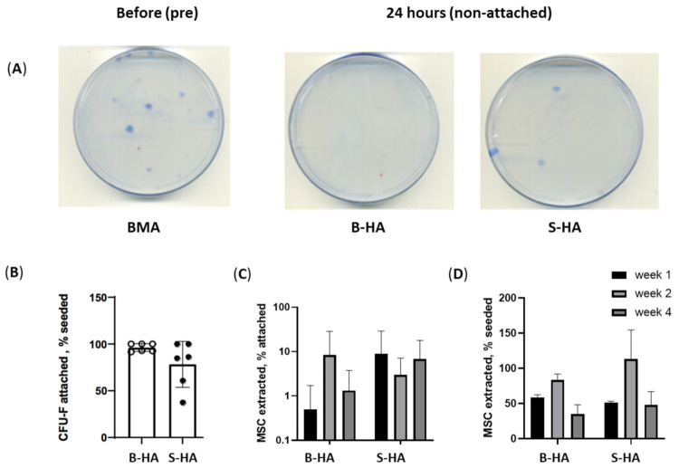
Figure 2.
BMSC and cMSC attachment and survival on the scaffolds. (A) Examples of BMSC colonies (CFU-F) following the plating of BMA without any scaffolds (left), BMSC colonies/CFU-F non-attached on B-HA (centre) and S-HA (right) after 24 h of adding BMA to the scaffolds. (B) BMSC attachment to scaffolds measured as the percentage of attached CFU-Fs to both scaffolds relative to seeded CFU-F; (C) BMSC maintenance measured as the percentage of BMSCs extracted after week 1, 2 and 4 of culture; (D) cMSC maintenance measured as the percentage of cMSCs extracted after week 1, 2 and 4 of culture using trypan blue for cell counts. Data are shown with mean and standard error of mean and with n = 3 cMSCs and n = 6 BMSCs.

