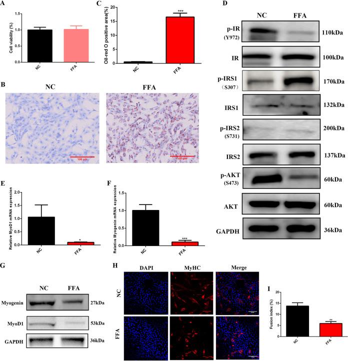
FIG 1.
Myogenic differentiation was suppressed by 1 mM FFA. (A) CCK-8 assay 24 h after 1 mM FFA treatment (n = 6). (B and C) Oil red staining 24 h after 1 mM FFA treatment (n = 3). (D) Western blotting of insulin signaling 24 h after 1 mM FFA treatment (n = 3). GAPDH was used as a loading control in all Western blot experiments. (E and F) Relative mRNA expression of MyoD1 and myogenin at 3 days of differentiation after 1 mM FFA treatment (n = 3). (G) Western blotting of MyoD1 and myogenin at 3 days of differentiation after 1 mM FFA treatment (n = 3). (H and I) Immunofluorescence staining of MyHC-positive cells at 3 days of differentiation after 1 mM FFA treatment (n = 3). *, P < 0.05 compared with NC; **, P < 0.01 compared with NC; ***, P < 0.001 compared with NC. Error bars indicate SD.

