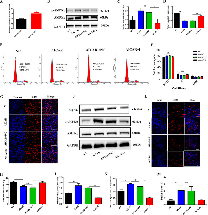
FIG 5.
AMPKα promoted myogenesis by arresting the cell cycle and decreasing cell proliferation through miR-206. (A) Relative mRNA expression of cyclin D1 after AMPKα shRNA transfection (n = 3). (B) Western blotting of AMPKα and p-AMPKα in C2C12 myoblasts 24 h after cotreatment with AICAR and the miR-206 inhibitor (AICAR+i) (n = 3). (C) Relative expression of miR-206 in C2C12 myoblasts 24 h after cotreatment with AICAR and the miR-206 inhibitor (n = 3). (D) Relative expression of cyclin D1 in C2C12 myoblasts 24 h after cotreatment with AICAR and the miR-206 inhibitor (n = 3). (E and F) Effect of cotreatment with AICAR and the miR-206 inhibitor on the cell cycle (n = 3). C2C12 cells were treated with AICAR and the miR-206 inhibitor, and the cell cycle was then analyzed by flow cytometry 24 h later. The percentage of cells in each phase of the cell cycle was calculated using Modfit32 software. (G and H) Effect of cotreatment with AICAR and the miR-206 inhibitor on cell proliferation (n = 3). C2C12 cells were cotreated with AICAR and the miR-206 inhibitor, and proliferating cells were then stained with EdU 24 h later. The nuclei were stained blue, and EdU-positive cells were stained red. The percentage of EdU-positive cells was calculated with IPP software. (I) Relative expression of miR-206 in C2C12 myotubes at 3 days of differentiation after cotreatment with AICAR and the miR-206 inhibitor (n = 3). (J and K) Western blotting of MyHC, AMPKα, and p-AMPKα in C2C12 myotubes at 3 days of differentiation after cotreatment with AICAR and the miR-206 inhibitor (n = 3). (L and M) Immunofluorescence staining of MyHC-positive cells at 3 days of differentiation after cotreatment with AICAR and the miR-206 inhibitor (n = 3). *, P < 0.05; **, P < 0.01; ***, P < 0.001; NS, not significant. Error bars indicate SD.

