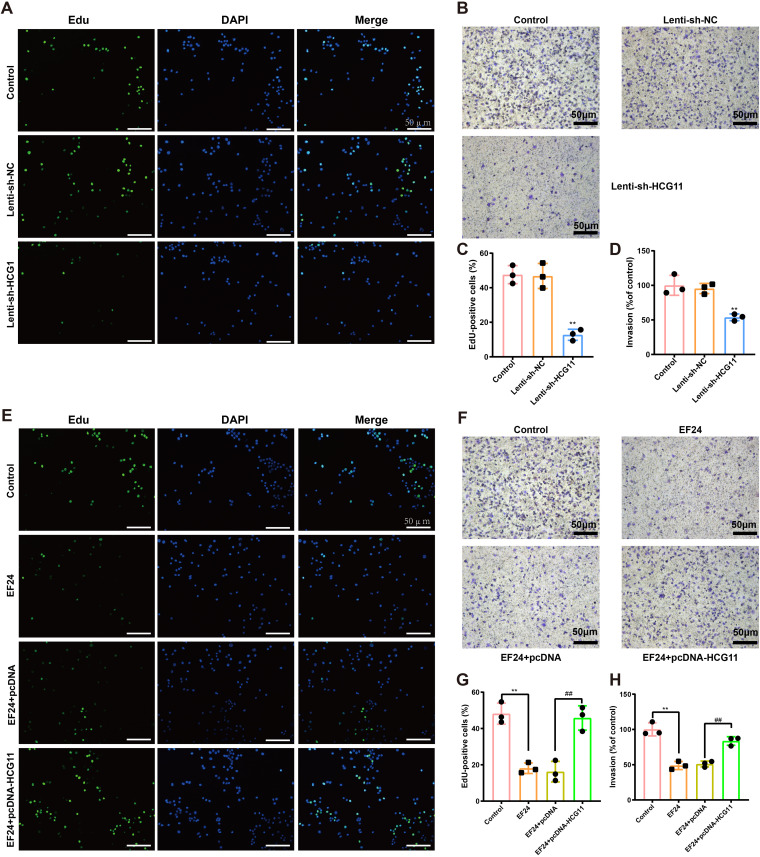
FIG 3.
HCG11 mediated the inhibitory effect of EF24 on cell proliferation and invasion in TNBC cells. (A to D) MDA-MB-231 cells were transfected with lentivirus (Lenti)-sh-HCG11 or its negative control (Lenti-sh-NC). Cell proliferation was measured using 5-ethynyl-2’-deoxyuridine (EdU) staining. Cell nuclei were counterstained with DAPI. Representative images (scale bar = 50 μm) were shown in panel A, and quantitative results were shown in panel C. Cell invasion was measured using Transwell assay. Representative images (scale bar = 50 μm) were shown in panel B and quantitative results were shown in panel D. (E to H) MDA-MB-231 cells were transfected with pcDNA-HCG11 or its negative control (pcDNA), followed by incubation of EF24. (E and G) Representative images (scale bar = 50 μm) (E) quantitative results (G) of EdU staining. (F and H) Representative images (scale bar = 50 μm) (F) quantitative results (H) of Transwell assay. Shapiro-Wilk test was used to check whether the data follow a normal distribution and F-test was used to verify if the variances were significantly different. F-test P < 0.05: Brown-Forsythe and Welch ANOVA test. F-test P ≥ 0.05: ordinary ANOVA test. (C and D) **, P < 0.01 versus Lenti-sh-NC; (G and H) **, P < 0.01; ##, P < 0.01.

