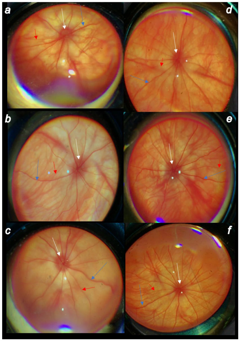
Figure 6.
Cauterization of the three episcleral veins induced posterior eye segment changes (veins (blue arrows), arteries (red arrows), optic disc (white arrows)) assessed by fundoscopy. Therapeutic regimen. (a). First post-injury day, regular course. (b). Fourth post-operative week, regular course. (c). Sixth post-injury week, regular course. (d). First post-injury day, course in BPC 157-treated rats. (b). Fourth post-operative week in BPC 157-treated rats. (c). Sixth post-injury week in BPC 157-treated rats. There were the discrete generalized irregularity in the diameter of the retinal vessels with mild atrophy of the optic disc, less visible normal presentation of the choroidal blood vessels at first post-injury day (a), progressively leading to evident worsening at the fourth post-operative week (b) with the strong generalized irregularity of the diameter of the blood vessels with severe atrophy of the optic disc and barely visible (extremely faint presentation) choroidal blood vessels (bright fundus background color) at the end of the sixth post-injury week (c). Contrarily, consequent to the evidence that all BPC 157 regimens, both prophylactic regimen and later, curative regimen, strongly reversed the increased intraocular pressure and abrogated mydriasis, illustrated are the normal fundus presentation, the presentation of the normal retinal and choroidal blood vessel presentation, and normal optic nerve presentation at the first post-operative day (d) (1 h after BPC 157 therapy application), fourth post-operative week (e) and sixth post-operative week (f) in the rats that started with BPC 157 therapy at post-surgery day 1. A similar effect was noted with all BPC 157 regimens. A total of 6 rats/group/interval.

