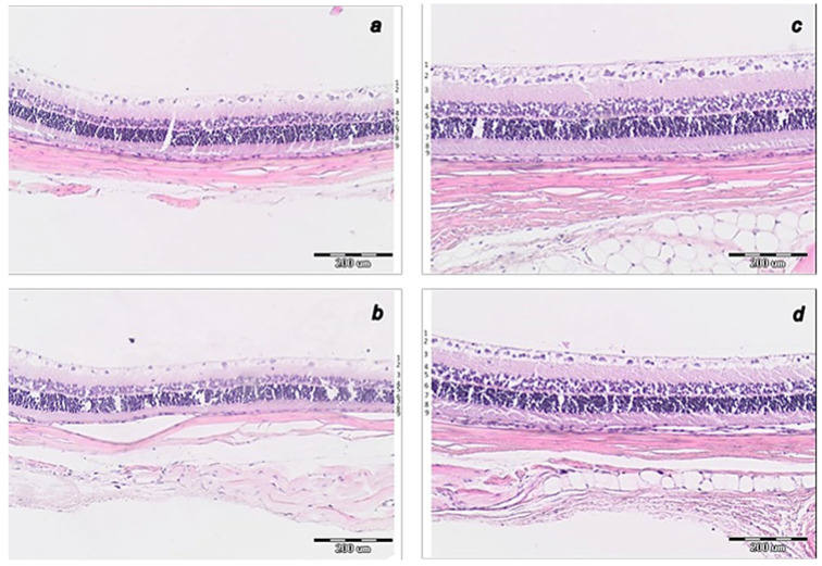
Figure 11.
Histology of central part of transverse section of the retina 0.4–0.7 mm on the temporal side of the optic disc. (a). Rat retina after episcleral vein cauterization at week 4 in controls. (b). Rat retina after episcleral vein cauterization at week 6 in controls. (c). Rat retina after episcleral vein cauterization at week 4 in BPC 157-treated rats. (d). Rat retina after episcleral vein cauterization at week 6 in BPC 157-treated rats. (HE staining, magnification ×20, scale bar 200 μm). Transverse section of the retina (0.4–0.7 mm on the temporal side of the optic disc) showing a strict difference in the retinal layers and full retina thickness in the rats that received saline and those that received BPC 157. More regular inner and outer nuclear layer and more regular distribution of ganglion cells, preserved thickness of retina, the inner and outer plexiform layer at week 4 in BPC 157-treated rats (c) than in control rats (a). At week 6, BPC 157-treated rats show the preserved thickness of the whole retina and also the inner plexiform layer and inner nuclear layer and its organization (d). Contrarily, degeneration of ganglion cells in control group is the most evident also as degeneration in inner and outer nuclear layer (b). Retinal layers as follows: 1—internal limiting membrane; 2—nerve fiber and ganglion cell layers; 3—inner plexiform layer; 4—inner nuclear layer; 5—outer plexiform layer; 6—outer nuclear layer; 7—outer limiting membrane; 8—photoreceptor layer; 9—pigment epithelium. A similar effect was noted with all BPC 157 regimens. A total of 6 rats/group/interval.

