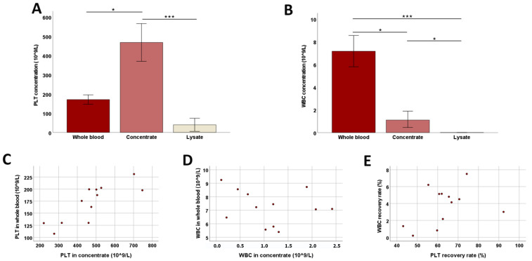
Figure 1.
Platelet and white blood cell counts during blood processing. Median platelet (PLT; (A)) and white blood cell (WBC; (B)) counts at the different stages of blood processing (whole blood, concentrate, and lysate before filtration) are presented; error bars display the 95% confidence intervals. Friedman tests for group comparisons and subsequent post hoc tests were performed; the asterisks describe the significant differences between groups (* corresponds to p < 0.05; *** corresponds to p < 0.001). The dot plots show PLT (C) and WBC (D) counts in whole blood vs. concentrate and WBC vs. PLT recovery rates (E); the correlation shown in (C) was significant (p < 0.001 and r = 0.820, based on Spearman’s rank correlation). All data were obtained from n = 12 dogs.

