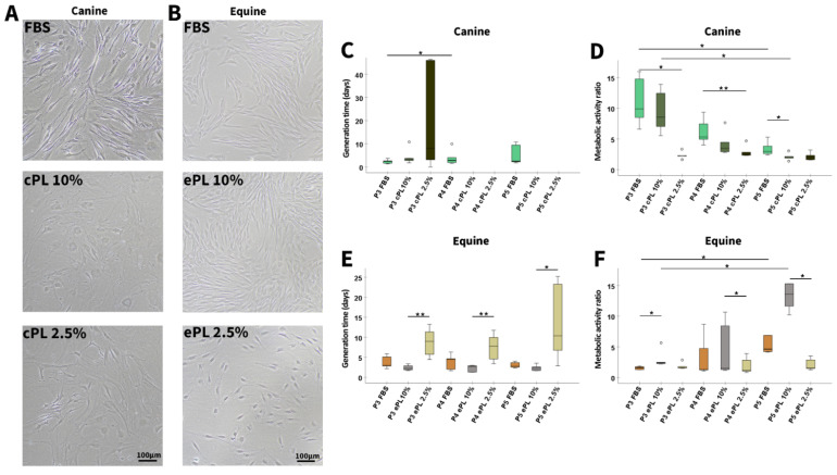
Figure 4.
Mesenchymal stromal cell culture with different media supplements. Representative phase-contrast photomicrographs show canine (A) and equine (B) mesenchymal stromal cells (MSC) in passage 4 and at day 5 after seeding for population doubling assays in media supplemented with fetal bovine serum (FBS) or canine/equine platelet lysate (cPL/ePL). The boxplots display the generation times calculated for canine (C) and equine (E) MSC, as well as their metabolic activity as determined by MTS tetrazolium-based cell proliferation assay for canine (D) and equine (F) MSC in passages 3 to 5 (P3 to P5); note that missing generation time data were due to insufficient proliferation in these samples. Friedman tests for group comparisons and subsequent post hoc tests were performed; the asterisks indicate the significant differences between the corresponding groups (* corresponds to p < 0.05; ** corresponds to p < 0.01). Data were obtained using MSC from the same n = 5 donor dogs or horses in each group.

