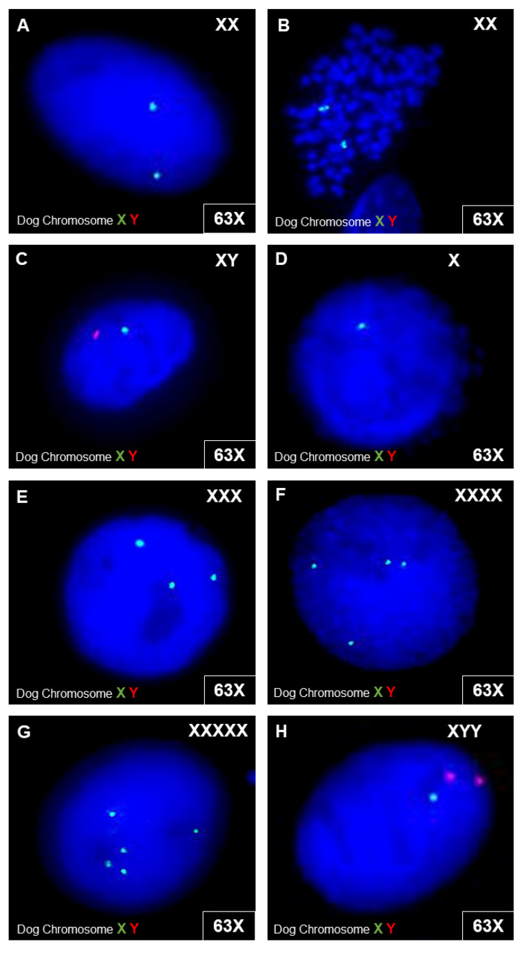
Figure 8.
Cytogenetic analyses in canine cells. FISH analyses were performed with dog chromosome XY FISH probes (centromeric alpha satellite DNA probe; chromosome X—spectrum green; chromosome Y—spectrum red) on interphase cells (A,C–F) and one metaphase cell (B). Exemplary images show the detected signal constellations. In (A–C), microscopic images of interphase cells and metaphase cells with a normal gonosomal karyotype are shown. In (D–H), examples of interphase cells with different signal patterns are shown, with one signal for X (D), three signals for X (E), four signals for X (F), and five signals for X (G). For some interphase cells, a combination of signals from one X-chromosome and two Y-chromosomes were detected (H).

