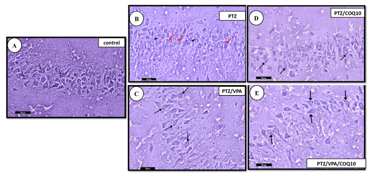
Figure 7.
Photomicrographs of the Toluidine blue-stained section of the hippocampus of all of the rat groups. (A) Control group showing that the cytoplasm of the pyramidal cells appears heavily studded with Nissl granules (↑). (B) PTZ group showing an apparent decrease in the Nissl granule content (red ↑) of the pyramidal cells of the pyramidal layer, compared to that of group I (control, non-epileptic rats). The pyramidal layer with few of the pyramidal cells has large vesicular nuclei (black ↑). (C) PTZ/VPA group showing an apparent increase in the Nissl granule content (↑). (D) PTZ/COQ10 group showing an apparent moderate increase in the Nissl granule content (↑) of the pyramidal cells of the pyramidal layer. (E) PTZ/VPA/COQ10 group showing an apparent increase in the Nissl granule content (↑). Toluidine blue; scale bar 50 µm.

