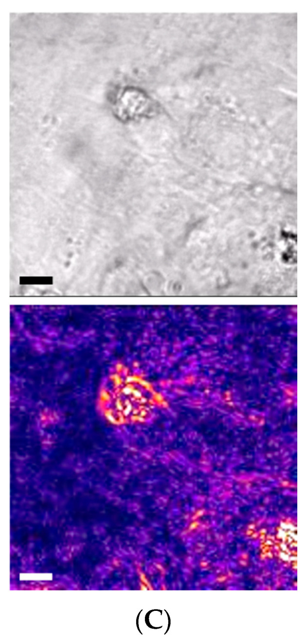
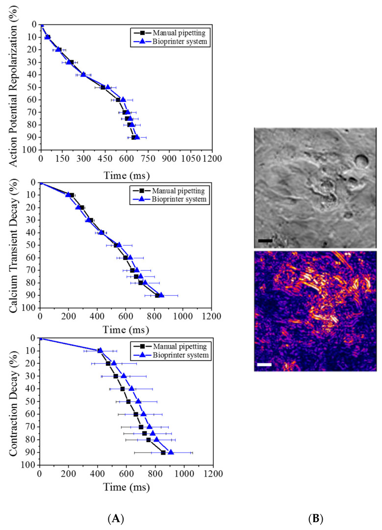
Figure 4.

Electrophysiological measurements of 2D tissue constructs delivered by manual pipette (control) or bioprinter valve (A). The three parameters assessed are the electrical activity on the cells (action potential), intracellular Ca2+ (calcium transient) and mechanical activity (contractile kinetic decay). Contractility images of printed cardiomyocytes, bright-field and computed contractility expressed as brightness: control cells deposited using pipette (B) and bioprinted cells (C), the scale bar represents 10 microns.

