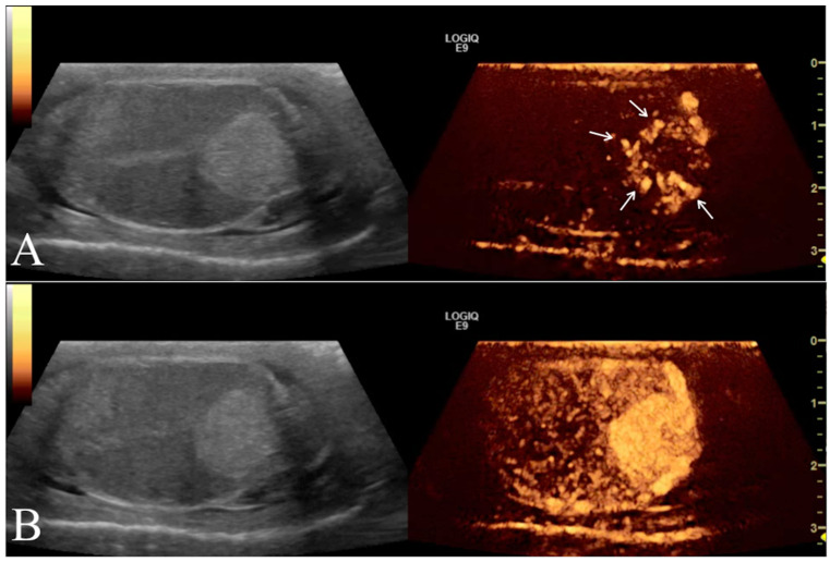
Figure 4.
Left testicle leydigoma of a 11 year old Italian Mastiff (the same of Figure 2). The left panel shows the sagittal B-mode ultrasound highlighting a focal hyperechoic nodule with homogeneous echotexture and regular margin. Representative contrast-enhanced ultrasound images of different contrast distribution phases are represented in the right panels. After 16 s from contrast injection it is possible to appreciate an early distribution of the contrast within the lesions (Arrows) compared to the surrounding parenchyma (A). Later (25 s) the contrast medium distributes homogeneously within the lesion that remain hyperenhanced compared to the normal testicle (B).

