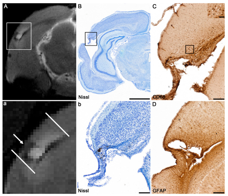
Figure 2.
Assessment of chronic neuroinflammation. (A) Magnetic resonance imaging (MRI) was used to identify animals with a prominent increase in T2-enhancement around the lesion (arrow in a high magnification representation (a)) indicating perilesional inflammation. (B) Nissl-stained sections show detailed cytoarchitectonics of the perilesional area. (C,D) Immunohistochemical stainings were used to confirm chronic neuroinflammation suggested by T2-enhancement. Immunohistochemical staining for CD68 (macrosialin) showed activated microglia/macrophages in the perilesional cortex (panel (C)). Immunohistochemical staining for glial fibrillary acidic protein (GFAP) with qualitative analysis showed astrocytic fibers (panel (D)). Scale bars = 2000 µm (B), 200 µm (b,C,D) and 50 µm (insert in panel (C)).

