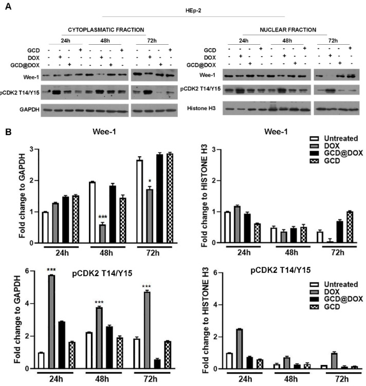
Figure 3.
Wee-1 and phospho-CDK2 (pCDK2 T14/Y15) protein expression in GCD@DOX-, GCD- and DOX-treated cells. HEp-2 cells were untreated or untreated with 25 μg/mL of GCD@DOX for 24 h, 48 h and 72 h. DOX (1.25 μg/mL) and GCD (25 μg/mL) were used as controls. (A) An equal amount of cytoplasmic and nuclear proteins was separated by polyacrylamide gel electrophoresis and probed with Wee-1 and phospho-CDK2 (pCDK2 T14/Y15) antibodies. GAPDH and Histone H3 were used as a loading control for the cytoplasmic and nuclear fractions, respectively. (B) The quantitative densitometric analysis of Wee-1 and pCDK2 T14/Y15 band intensities was determined in the cytoplasmatic and nuclear fractions for both with ImageJ software and expressed as fold change over the appropriate housekeeping gene. (* p < 0.1; *** p < 0.001).

