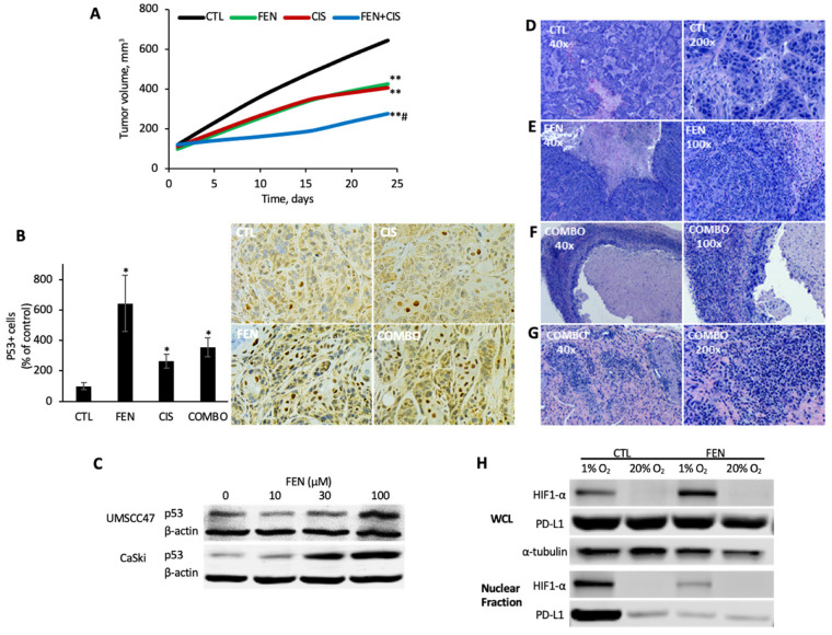
Figure 6.
Fenofibrate inhibits in vivo tumorigenicity, promotes p53 accumulation, and reprograms the tumor-immune microenvironment. (A) UMSCC47 xenograft model. UMSCC47 cells were implanted into the flanks of athymic nude mice and tumors were allowed to develop without intervention. Tumor-bearing mice (~100 cm3) were randomized to four treatment arms: control (CTL), fenofibrate (FEN; 200 mg/kg PO, 5 times/week), cisplatin (CIS; 2 mg/kg IP; once weekly), or fenofibrate + cisplatin (COMBO). ** FEN or CIS vs. CTL, p < 0.001 or COMBO vs. CTL, p = 0.0001; # COMBO vs. FENO or CIS, p = 0.0001. (B) p53 immunohistochemistry. p53 levels were analyzed in harvested UMSCC47 xenograft tumors and tumor cells with strong nuclear staining was scored as p53+. A representative IHC image for each experimental condition is shown (200×). * p < 0.01. (C) p53 levels. Immunoblot for p53 protein levels following control or fenofibrate treatment in UMSCC47 cells. Uncropped immunoblots are available in File S1. (D–G) Hematoxylin- and eosin-stained tumor sections. Representative tumor sections illustrating notable morphological features, at low power (40×; left) and higher power magnification (100–200×; right). (H) HIF1α and PD-L1 levels. Immunoblot for nuclear HIF1α and PD-L1 levels under normoxic (20% O2) or hypoxic (1% O2) conditions with control or fenofibrate treatment in UMSCC47 cells. Uncropped immunoblots are available in File S1.

