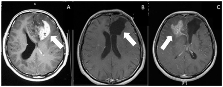
Figure 1.
Post gadolinium contrast administration, T1-weighted axial images. (A) Preoperative, heterogeneous irregular enhancement, associated with the left frontal-lobe glioblastoma (arrow). (B) Postoperative (at 1 month) axial weighted image. On postoperative image, there is no residual enhancement. Arrow shows operation cavity. (C) Postoperative (at 18 months) axial weighted image shows recurrence of the tumor (white arrow) on contralateral hemisphere, associated with peripheral edema.

