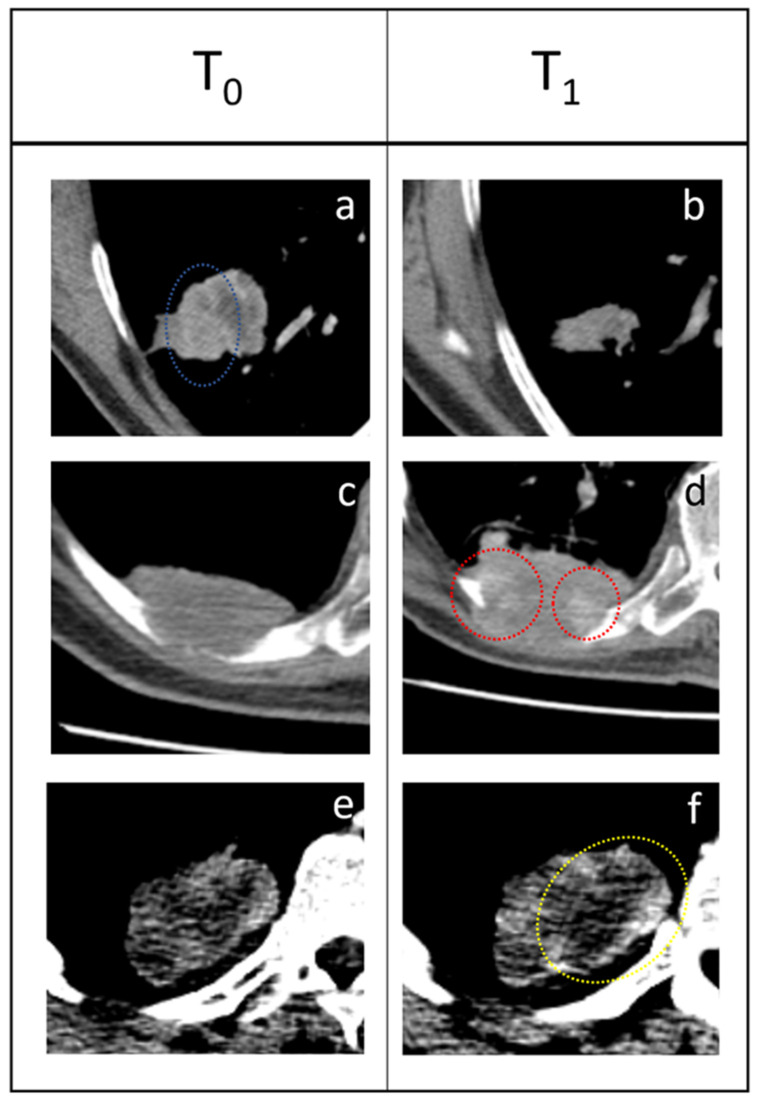
Figure 3.
Three examples of response to immunotherapy. (a,b) PR: the tumor shrunk from T0 to T1 and areas that presented high enhancement (blue dotted circle) disappeared. (c,d) SD: 63-year-old male patient presenting a pulmonary tumor that penetrates the thoracic wall and erodes a rib with extensive bone reabsorption. The first re-evaluation showed an increased mass (53 × 35 mm vs. 50 × 25 mm) consistent with PD. However, during therapy, the lesion gradually reduced and became stable. Delta-radiomics individuated an extreme negative variation of LAHGLE (−0.999770105) that could be represented by the emergence of areas with vivid contrast media uptake (red dotted circles) within the lesion. (e,f) PD: this pulmonary lesion was slightly enlarged at the first follow-up but radiomic revealed an increase in LALGLE (−0.173288587). The lesion kept growing in the following months, a finding consistent with PD. This case represents a good example of how radiomics could intercept changes in the radiological images that are almost invisible to the human eye and predict the evolution of each lesion. The slight increase of LALGLE could be referred to as the slight enlargement of the central hypodense area (yellow dotted circle). In this case, the CT window was stressed for demonstrative purposes.

