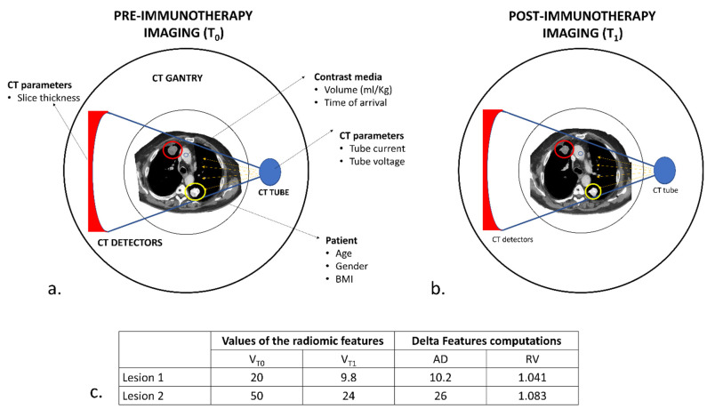
Figure 7.
Hypothetical schematic representation of the variability in CT acquisition and delta-radiomic computations. (a) Baseline CT (T0) in a patient with two pulmonary lesions. Several factors might influence CT acquisition, depending on the type of scanner (number and dimensions of the detectors), the acquisition parameters employed (tube current and voltage) and the characteristics of the patient (age, gender and body mass index) from which depends, to some extent, the contrast medium kinetic and distribution. Even the position within the body of the pulmonary lesion might affect the radiomic analysis, as the values of the pixels depending on the attenuation of x-rays. Lesion 2 (yellow circle) lies between two high-attenuated bony structures (*), the spine and the scapula; lesion 1 (red circle), on the other hand, lies nearer to the skin and distant from the ribs or sternum. An x-ray beam will interact with these two lesions differently, resulting in different values of radiomic features. (b) At the first re-evaluation (T1), the two lesions responded to immunotherapy. In this case, each lesion will be acquired in conditions very similar to T0. (c) During delta-radiomic features computation, variations due to CT acquisition are eliminated using the RV method, which can extrapolate a similar change in different situations. We can appreciate how the two lesions, despite their differences in values and AD-calculated delta features, presented comparable changes.

