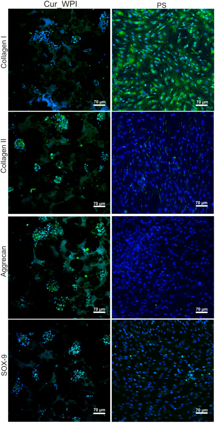
Figure 8.
Confocal microscope images presenting characteristic markers—collagen II, aggrecan, and SOX-9 after the 12-day culturing of chondrocytes on Cur_WPI biomaterials and polystyrene (PS). Collagen I was also visualized to determine chondrocyte dedifferentiation towards fibroblast-like cells. Nuclei emitted blue fluorescence, while evaluated markers gave green fluorescence (visible green fluorescence in the structure of biomaterial was emitted by WPI); magnification 200×, scale bar equals 70 μm.

