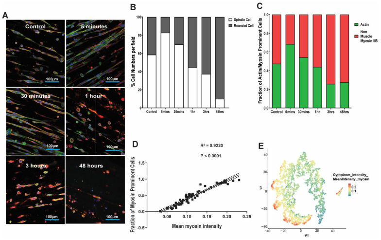
Figure 5.
Kinetics of lung fibroblast cytoskeleton and morphological alterations following mechanical strain. Human fetal lung 1 (HFL1) fibroblasts were embedded in 3D collagen-1 gels of 2.0 mg/mL concentrations and cultured for 24 h. The HFL1 fibroblast-seeded collagen gels were then left alone or mechanically strained for 48 h at a 1% amplitude and a frequency of 0.2 Hz. Gels were collected after 0, 5, and 30 min, and 1, 3, and 48 h, following the stain protocol. (A) Representative confocal images of HFL1 fibroblasts in 2.0 mg/mL collagen-1 gels stained for non-muscle myosin IIB (red) and F-actin (green) after mechanical strain experiments at different time points. (B) Percentage cell numbers of spindle and rounded shaped cells at different time points in 2.0 mg/mL collagen-1 gels after being strained at different time points. (C) Percentage positive pixel count per total number of pixels in images of 2.0 mg/mL collagen-1 gels after being strained at different time points. (D) The correlation between the fraction of myosin prominent cells and the mean myosin intensity in images of 2.0 mg/mL collagen-1 gels after being strained at different time points. (E) TSNE-plot visualization of each single cell with the 2.0 mg/mL collagen-1 gels after being strained at different time points. Mean stacked bar graphs are shown for 3 technical replicates, n = 6 in (B,C).

