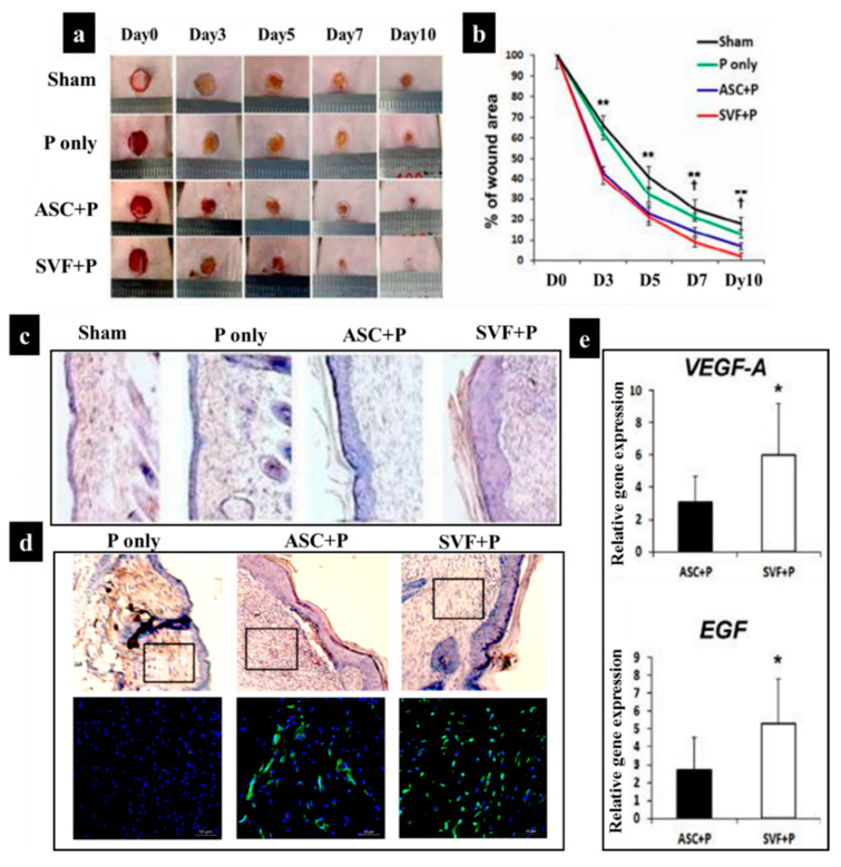
Figure 6.
(a) Wound contraction over a 10-day period in four groups: sham, pluronic hydrogel (P) only, ASC+P, and SVF+P; (b) diagram showing the percentage of wound area each day; (c) histological images after H&E staining at 14 days after the injection of cells. (d) Representative photographs of isolectin B4 (ILB4), which is a marker of endothelial cells for neovascularization, in the skin wounds at 14 days after cell transplantation; nuclear DAPI staining is blue and ILB4 staining is green. On the upper line of the picture are the results of H&E from wound tissue after cell injection. The black rectangular box indicates the location of endothelial cells that show capillary density. (e) Quantification of expression levels for VEGF-A and EGF in skin wound tissues (* p < 0.05, ** p < 0.01) [162].

