Figure 2.
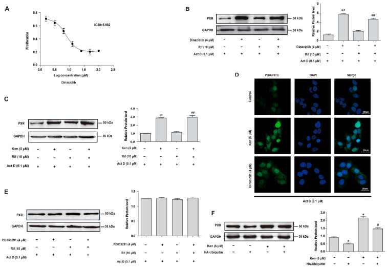
Inhibition of CDKs increases the protein level of PXR by suppressing its ubiquitination. (A) The cytotoxicity of dinaciclib in HepG2 cells. Hepatocytes were treated with indicated concentrations of dinaciclib for 24 h, and MTT assay was used to measure cell viability. (B–E) The effect of CDKs inhibitors on PXR protein stabilization in HepG2 cells. (B) Cells were treated with dinaciclib (4 μM) in the presence of actinomycin D (0.1 μM) with or without rifampicin (10 μM) for 24 h. (C) HepG2 cells were treated with kenpaullone (Ken, 5 μM) in the presence of actinomycin D (0.1 μM) with or without rifampicin (10 μM) for 24 h. (D) HepG2 cells were treated with kenpaullone (5 μM) or dinaciclib (4 μM) in the presence of actinomycin D (0.1 μM) for 24 h, and fluorescence intensity of PXR was detected by immunofluorescence analysis. (E) HepG2 cells were treated with PD033291 (4 μM) in the presence of actinomycin D (0.1 μM) with or without rifampicin (10 μM) for 24 h. PXR protein levels were investigated by Western blot. (F) HepG2 cells were transfected with HA-ubiquitin for 24 h and then treated with or without kenpaullone (5 μM) for 24 h. PXR protein levels were investigated by Western blot. Experiments described in this figure were repeated independently at least three times, and data are expressed as mean ± SEM (n = 3). * p < 0.05, ** p < 0.01 versus control; # p < 0.05 versus HA-ubiquitin overexpression group, ## p < 0.01 versus rifampicin-treated group.

