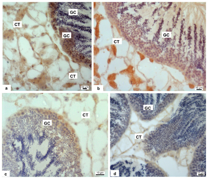
Figure 3.
Immunolabeling of M. galloprovincialis male gonad with anti-3β-HSD antibody. Brown coloration indicates the enzyme presence. Nonexposed (a) and exposed mussels to 1 (b), 10 (c) and 100 pM (d) HgCl2, respectively. Germ cells (GC) are positive to antibody in control and 1 and 10 pM treated samples (a–c); the positivity disappears almost completely in 100 pM treated samples (d) In the connective cells (CT, connective tissue) the 3β-HSD enzyme is recognizable in control and 1 pM treated animals (a,b); in 10 and 100 pM treated samples the antibody positivity is poor (c) or totally disappeared (d).

