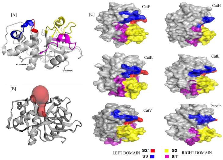
Figure 1.
(A) Cartoon representation of Cathepsin with Left (L) and Right (R) domains consisting substrate binding subsites S2′ (red), S3 (blue) and S2(yellow), S1′(pink) respectively. The N-terminal L-domain contains mostly α-helix whereas the C-terminal R-domain with mostly β-sheets. The image was prepared by PyMOL. (B) The cartoon structure of protein is showing the position of active site cleft at the mid-point of L- and R-domain. The red spheres indicating the presence of binding pocket at this site. The image was created by Computed Atlas of Structure Topography of proteins (CASTp). (C) Surface structures of all the cathepsins are showing their structural similarity with papain, comprising S2′(red), S3(blue) substrate binding sites at Left (L) and S2 (yellow), S1′ (pink) substrate binding sites at Right (R) domains. All the images were created by using PyMOL.

