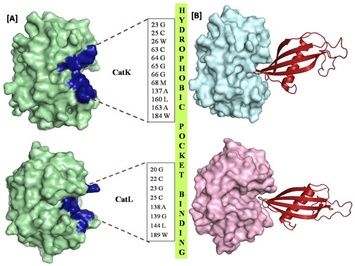
Figure 3.
(A) Representing the interacting residues of CATK in CYAN and CATL in PALEGREEN with CysC in MAGENTA forming hydrogen bonding network at the binding pocket. H-bonding could be observed between Tyr67 and Val83, Cys25 and Ala84 of CATK and Cys C, respectively, Asp162 and Val83, Trp189 and Trp132 of CATL and CysC respectively. (B) Representing the hydrophobic interacting residues of CATK and CATL with CysC. The binding of the CysC in the exosite of CatL is substantiated by the distribution of the hydrophobic moieties in the S2 and S1 subsites, whereas, CatK demonstrates a more uniform distribution the hydrophobic residues in the active site.

