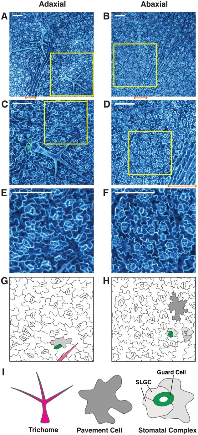
Figure 1.

Arabidopsis leaf epidermis and cell types. A–F, Representative images of the adaxial (A), (C), and (E) and abaxial (B), (D), and (F) epidermis of the third true leaf of 2-week-old Arabidopsis seedlings. Maximum fluorescence intensity projections of confocal z-stacks of the endoplasmic reticulum marker line 35S:GFP-HDEL (Haseloff et al., 1997) are shown for cell visualization. Scale bars represent 100 µm. The images in (C–F) correspond to the yellow insets in (A–D), respectively. G and H, Drawings of the cell outlines shown in (E) and (F), respectively, with representative cell types false colored to match with (I). Green asterisks in (C) indicate representative socket cells. Orange double arrows in (A), (B), and (D) indicate the location of a midvein, where pavement cells are rectangular and stomatal development is restricted. I, Representative drawings of trichome (magenta), pavement cell (gray), and stoma with paired guard cells (green)/stomatal complex (with SLGCs light gray). SLGCs are arranged in a spiral manner to form an anisocytic stomatal complex.
