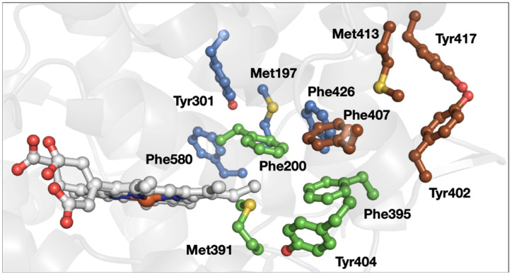
Figure 7.
Structure of prostaglandin H2 synthase 1 (PDB ID 1Q4G [54]). The 3-bridge clusters are highlighted in maroon, green, and lavender, and the heme is shown in gray. Red corresponds to oxygen, yellow to sulfur, and blue to nitrogen. The image was generated using PyMOL.

