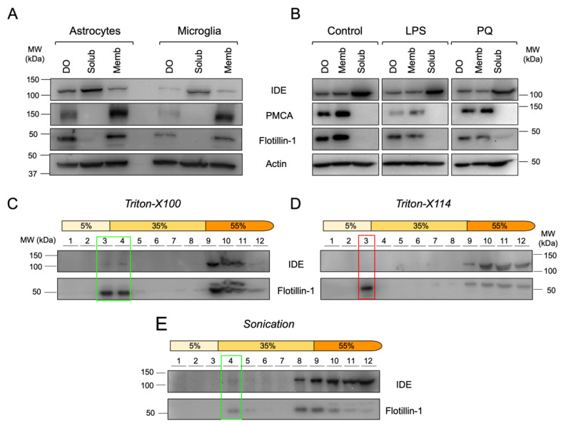
Figure 5.
IDE is stably associated to membranes of primary glial cells and to membrane microdomains with specific physicochemical properties in the microglial cell line BV-2. (A) Immunoblot analyses of primary glial cells upon centrifugal fractionation in dense organelles (DO), membrane (Memb) and soluble (Solub) fractions. (B) Immunoblot analyses of BV-2 microglial cells treated with different stimuli (100 ng/mL LPS or 25 μM PQ for 24 h) and fractionated in DO, Memb and Solub fractions. (C–E) Immunoblot analyses of membrane fractionation into lipid rafts and non-raft domains using different methods: Triton-X100 (C), Triton-X114 (D) and sonication (E). Flot-1 was used as a lipid raft marker. Rectangles highlight the lipid raft fractions.

