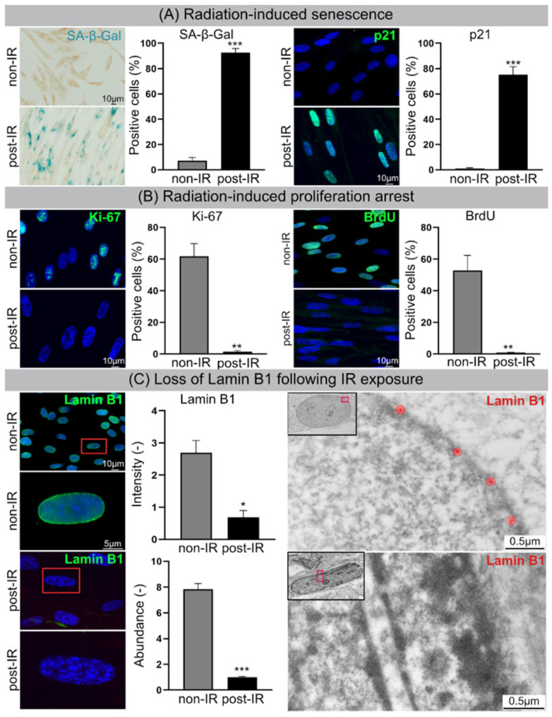
Figure 1.
Cellular senescence following IR. (A) Increased numbers of SA-β-Gal-positive and p21-positive cells following IR. (B) Decrease in Ki-67-positive and BrdU-positive cells. (C) Lamin B1 loss in nuclear envelope following IR exposure visualized by IFM (left) and TEM (right). Quantification of lamin B1 in WI-38 fibroblasts by IFM (top middle), and MS (bottom middle). Data are presented as mean ± SEM, * p < 0.05, ** p < 0.01, *** p < 0.001.

