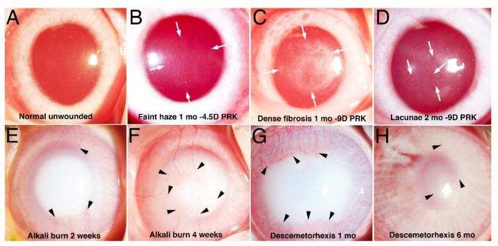
Figure 1.
Slit lamp photographs of haze and scarring fibrosis in rabbit corneas. (A) Normal unwounded transparent cornea. (B) One month after −4.5D PRK a cornea has faint opacity (haze) within arrows [5]. (C) One month after −9D PRK a cornea has dense scarring fibrosis within arrows [5]. (D) At 2 mo. after −9D PRK areas of clearing (lacunae, arrows) are developing within scarring fibrosis [5]. (E) Dense scarring fibrosis 2 weeks after 5 mm surface alkali burn with 1 N NaOH. Stromal neovascularization (arrowheads) begins to develop. (F) Scarring fibrosis has progressed at 4 weeks after alkali burn. Stromal neovascularization (arrowheads) has progressed. (G) Dense scarring fibrosis 1 mo. after 8mm Descemetorhexis. Stromal neovascularization (arrowheads) has developed [7]. (H) Scarring fibrosis has diminished by 6 mo. after Descemetorhexis with iris details now visible. Most of the opacity that remains is associated with the corneal neovascularization (arrowheads) [7]. Mag. 20×.

