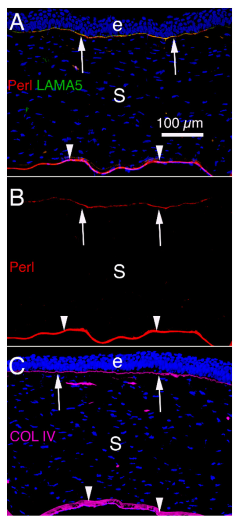
Figure 2.

Corneal BM components that modulate TGF beta-driven myofibroblast development and fibrosis in unwounded rabbit corneas [5]. (A) Immunohistochemistry (IHC) for perlecan (Perl), as well as laminin alpha-5 (LAMA5) [5]. (B) IHC for perlecan alone. (C) IHC for collagen type IV. Arrows indicate the EBM with overlying epithelium (e) and arrowheads indicate Descemet’s membrane that overlies the corneal endothelium, respectively, in all panels. S is stroma populated primarily with keratocytes. Blue is DAPI stained nuclei. Mag. 200×.
