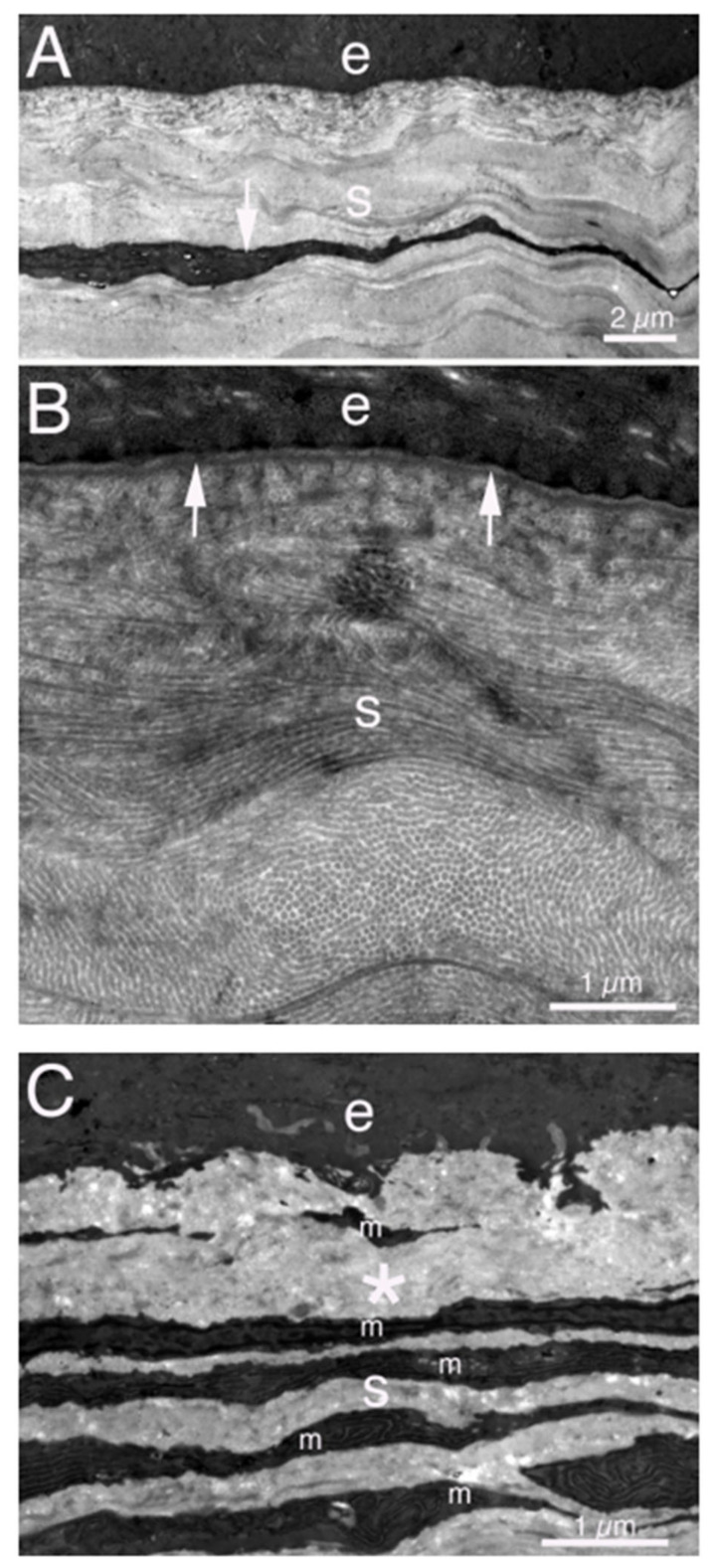
Figure 3.
TEM of normal and fibrotic rabbit corneas. (A) Lower magnification image of an unwounded cornea showing the epithelium (e) and stroma (s) with a keratocyte (arrow). (B) Higher magnification image showing the epithelium (e) with the underlying EBM. The arrows indicate the lamina lucida anterior to the lamina densa of the EBM. In the stroma (s) note the uniform diameter of the collagen fibrils, with some seen in cross-section and others longitudinally, and the highly ordered packing of the fibrils. (C) In a cornea with severe fibrosis at 1 month after PRK, the stromal ECM is highly disorganized (*), without evidence of regular fibrils or packing. The anterior stroma (S) is also populated with many layered myofibroblasts (m). These images were previously unpublished but from the study of Torricelli et al., Investig. Ophthalmol. Vis. Sci. 2013, 54, 4026–4033.

