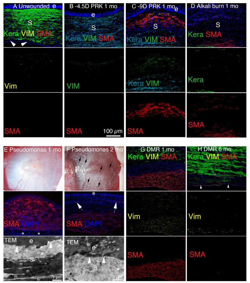
Figure 6.
Stromal cellularity of a normal cornea and corneas after injuries that heal without fibrosis and with fibrosis in rabbits. (A) The unwounded corneal stroma (s) is populated with keratocan-positive keratocytes between the epithelium (e) and corneal endothelium (arrowheads). At the vimentin (vim) antibody concentration used [5,25], only a few anterior stromal keratocytes were vimentin positive. No SMA-positive cells were detected. (B) One month after −4.5D PRK, there were numerous vimentin-positive corneal fibroblasts in the anterior stroma but the stroma was mostly populated with keratocan-positive keratocytes. No SMA-positive myofibroblasts were detected. (C) One month after −9D PRK, the anterior stroma is populated with SMA-positive, vimentin-positive myofibroblasts and SMA-negative, vimentin-positive corneal fibroblasts (and possibly fibrocyte progeny). (D) At 1 month after a one-minute 1N NaOH alkali burn that also destroyed the endothelium and Descemet’s membrane, the full-thickness corneal stroma is filled with myofibroblasts and corneal fibroblasts. Few keratocytes are detected. (E) At 1 month after infection with Pseudomonas aeruginosa keratitis sterilized with topical tobramycin there is severe opacity of the cornea in a slit lamp photograph. In IHC, approximately 90% of the stroma is filled with SMA-positive myofibroblasts, and in this cornea sparred only the most posterior stroma adjacent to the corneal endothelium. In a TEM image of this cornea, no lamina lucida/lamina densa is detected. The stroma (s) has disorganized ECM (*) and numerous myofibroblasts (m). (F) At 1 month after infection with Pseudomonas aeruginosa keratitis sterilized with topical tobramycin, the opacity in the cornea decreases and numerous transparent areas called lacunae (black arrows) develop. On IHC in this cornea where the Pseudomonas aeruginosa extended through the entire cornea and destroyed the corneal endothelium, SMA-positive myofibroblasts populate the posterior stroma but myofibroblasts disappeared in the anterior stroma. Corneal neovascularization (arrows) with SMA-positive pericytes develops. In a TEM image, lamina lucida/lamina densa (arrowhead) was regenerated. The stroma (s) had organized collagen fibrils similar to normal unwounded stroma and no myofibroblasts were detected in the anterior stroma. (G) At 1 month after Descemetorhexis (DMR), the posterior stroma is filled with SMA-positive myofibroblasts. The more anterior stroma in this section had keratocan-positive keratocytes. An intermediate layer of SMA-negative, keratocan-negative, vimentin-positive corneal fibroblasts (and likely fibrocyte progeny) are present between the keratocyte and myofibroblast layers. (H) At 6 months after DMR, the corneal endothelium (arrowheads) regenerates. Most of the posterior stroma is repopulated with keratocan-positive keratocytes, except adjacent to the corneal endothelium and regenerated DBM there were numerous keratocan-negative, SMA-negative, vimentin-positive corneal fibroblasts and a few remaining SMA-positive myofibroblasts. e is epithelium and s is stroma in all panels. Blue is DAPI-stained nuclei in all panels. Panels B and C reprinted with permission from de Oliveira et al. Exp Eye Res 2021:202;108325. Panels E and F reprinted with permission from Marino et al. Exp Eye Res. 2017;161:101-105. Panels G and H reprinted with permission from Sampaio LP et al. Exp Eye Res. 2021;213:108803.

