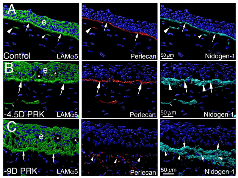
Figure 7.
Defective perlecan EBM incorporation in a PRK injured rabbit cornea that developed scarring stromal fibrosis and myofibroblasts. Confocal microscopy Imaris 3D constructed images of triplex IHC for laminin alpha-5, perlecan and nidogen-1 in an unwounded control cornea and corneas with moderate −4.5D PRK and severe −9D PRK epithelial-stromal injury [5]. (A) Laminin alpha-5 (green) was detected in the epithelium (e) and in the EBM (arrows) in an unwounded cornea. Two DAPI-negative vesicles with laminin alpha-5 (arrowheads) are present in the anterior stroma adjacent to the EBM. These were likely produced by keratocytes to contribute to maintenance of the EBM. Perlecan (red) was detected in the EBM (arrows), and in vesicles in the anterior stroma (arrowhead). Nidogen-1 (blue gray) is a major component in the EBM (arrows) and is present in secretory vesicles in the anterior stroma (arrowheads). (B) A cornea at 1 month after surgery that had moderate epithelial-stromal injury (−4.5D PRK) and did not develop myofibroblasts or scarring stromal fibrosis (see Figure 1B). The laminin alpha-5, perlecan and nidogen-1 localization in the EBM are similar to that noted in the unwounded cornea (large arrows), except there are increased nidogen-1 (arrowheads) in the subepithelial stroma surrounding stromal keratocyte/corneal fibroblast cells. Vesicles (small arrows) that are DAPI-negative are present in the anterior stroma and contain one or more of the EBM components. (C) In a cornea 1 month after more severe epithelial-stromal injury (−9D PRK) there is greater stromal opacity and myofibroblasts (see Figure 1C). Laminin alpha-5 and nidogen-1 (arrows) EBM localization is similar to that noted in the unwounded control cornea. Perlecan, however, was not detected at significant levels in the EBM, even though it is present within and surrounding myofibroblasts (arrowheads) in the anterior stroma. Stromal nidogen-1 (arrowheads) surrounding myofibroblasts is also present at high levels in the anterior stroma. Blue in all panels is DAPI-stained nuclei. e is epithelium. * indicates artifactual defects in the epithelium which are often noted in PRK corneas that are cryo-sectioned in the first 1 to 2 months after surgery while the EBM has not fully regenerated. Reprinted with permission from de Oliveira et al. Exp Eye Res 2021:202;108325.

