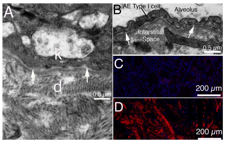
Figure 8.
Organs where BMs can have a role in fibrosis [43]. (A) TEM in normal rabbit skin. The basal keratinocyte (k) and dermis (d) are separated by the BM with lamina lucida (arrows) and lamina densa. Note the larger and more disorganized fibrils in the dermis compared with corneal stroma in Figure 3b. (B) TEM in normal rabbit lung. The alveolar BM with lamina lucida (arrow) and underlying lamina densa separates the alveolar epithelial cell type I (AE cell type I) from the interstitial space. (C) IHC for SMA in normal human lung primarily stains pericytes associated with blood vessels. There is little staining for SMA in the normal lung parenchyma. Blue is DAPI stained nuclei. (D) In a human lung with advanced idiopathic pulmonary fibrosis (IPF) SMA-positive myofibroblasts are present throughout the parenchyma. Blue is DAPI stained nuclei.

